

This article gently guides you through the preparation and insights related to cardiology PET scans, emphasizing their vital role in diagnosing heart conditions. Understanding that you may have concerns, we highlight the necessary steps you can take to prepare for the procedure with confidence. These scans are essential for detecting issues like coronary artery disease and assessing heart function.
In addition to this, we provide detailed guidelines on:
Our aim is to ensure that you feel supported and informed throughout this process, leading to a successful experience. Remember, taking these steps not only aids in your diagnosis but also contributes to your overall well-being.
A cardiology PET scan is a vital tool in modern heart diagnostics, providing a non-invasive look into the complex workings of the heart. This advanced imaging technique not only helps identify coronary artery disease but also assesses the effectiveness of treatments. Such capabilities make it invaluable for patient care, especially for our elderly loved ones.
However, the path to obtaining accurate results can sometimes feel overwhelming, filled with uncertainties and preparation challenges. How can patients navigate the complexities of a cardiac PET scan to ensure they receive the most accurate diagnosis and optimal care?
By understanding the process and preparing adequately, patients can take control of their health journey. It's essential to feel supported and informed every step of the way, ensuring that the experience is as smooth and reassuring as possible.
A cardiology PET scan (Positron Emission Tomography) is a non-invasive imaging test that provides high-resolution images of the heart's structure and function. By using a small quantity of a radioactive substance known as a tracer, this process helps us understand how effectively blood circulates to the cardiac muscle. The emitted positrons from the tracer are captured by the PET scanner, creating detailed images that are crucial for diagnosing various heart conditions. Importantly, PET/CT imaging of the heart involves very low radiation exposure, making it a safe choice for older patients.
The significance of cardiology PET scan is highlighted by its ability to:
This imaging technique is especially beneficial for elderly patients, who often present with atypical symptoms or have multiple health issues that complicate diagnosis. Recent advancements in cardiology PET scan imaging technology, particularly the N-13 Ammonia Heart PET/CT offered by Amavita Heart and Vascular Health®, have further enhanced its diagnostic capabilities. This allows for the detection of heart disease years before traditional methods, often when treatment is most effective and outcomes are optimal.
Typically, a cardiology PET scan is completed in around 30 minutes, providing valuable diagnostic insights. Statistics show that coronary artery disease is prevalent among older adults, making accurate and timely diagnosis essential. Real-world examples illustrate how heart PET imaging has effectively identified coronary artery illness in individuals who might have otherwise gone unnoticed. As Dr. Marcelo Di Carli, a professor of radiology and medicine at Harvard Medical School, states, '[PET/CT] helps us differentiate a patient who has chest pain from obstructions of the coronary artery from another patient who may also have chest pain, but not coming from obstructed coronary arteries.' This ability to is invaluable, particularly for those with complex health profiles. Furthermore, recent guidelines recommend PET over other modalities for quantitative myocardial blood flow evaluation, highlighting its growing importance in heart diagnostics.
If you or a loved one are experiencing concerns about heart health, consider discussing the benefits of cardiac PET imaging with your healthcare provider. Your health is paramount, and understanding these options can lead to better outcomes and peace of mind.

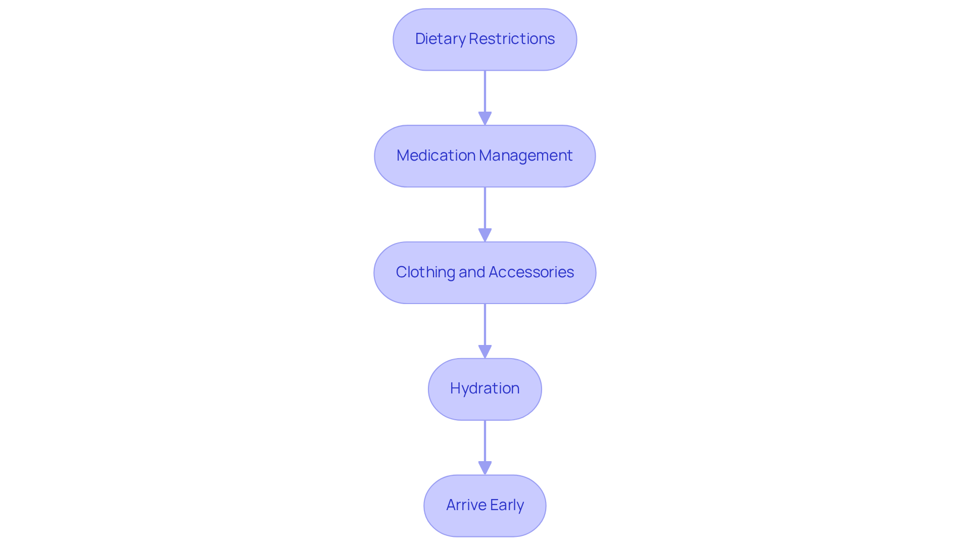
To ensure a successful cardiac PET scan, it's important to follow some essential guidelines that can help ease your mind and prepare you for the procedure:
By following these guidelines, you can feel more prepared and at ease for your cardiology PET scan. Remember, your healthcare team is here to support you every step of the way.

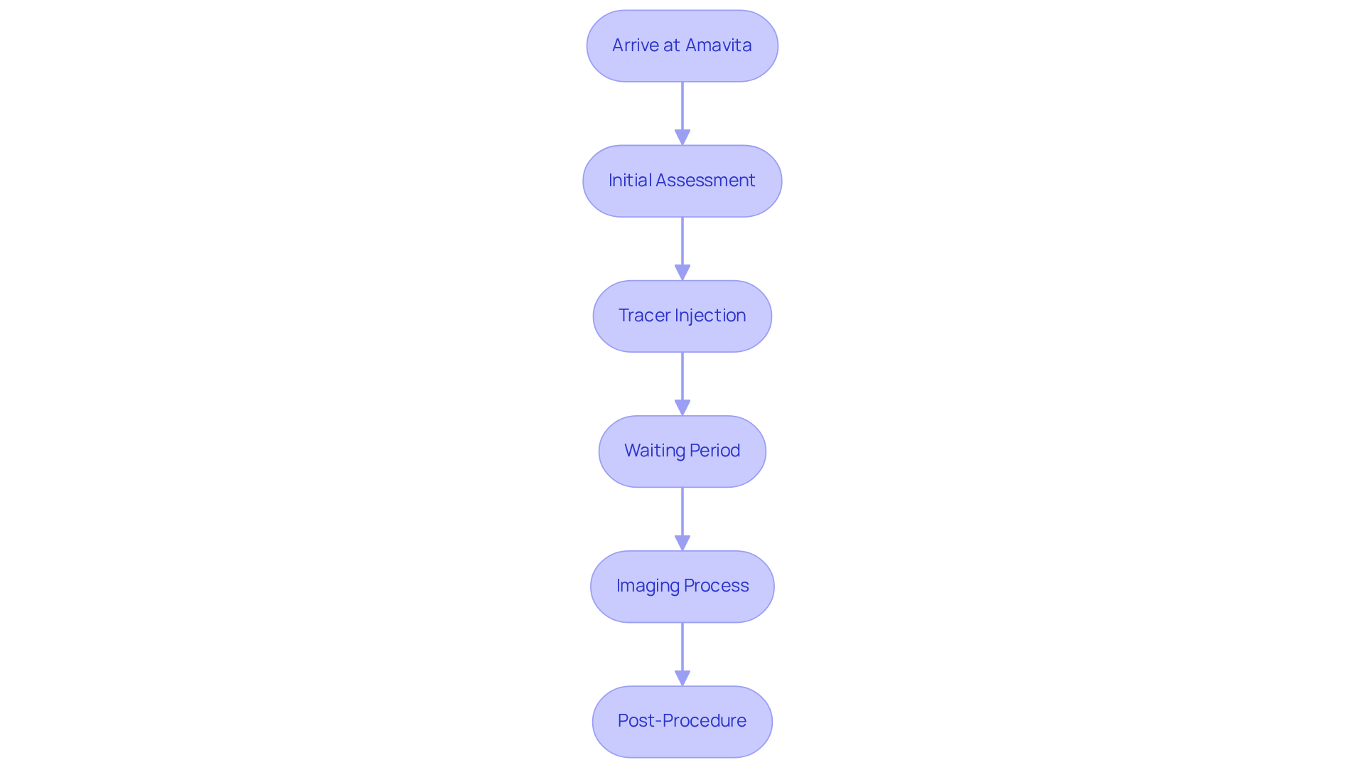
At Amavita Heart and Vascular Health, we understand that undergoing a cardiology PET scan can be a source of anxiety for many patients. Here’s what you can expect during your visit:

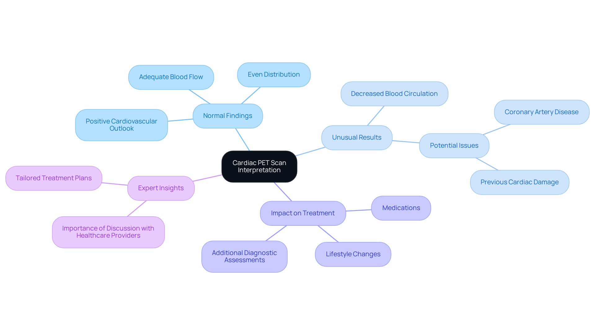
Interpreting the results of your cardiac PET scan involves understanding some important aspects that can affect your health journey:

Understanding the intricacies of a cardiology PET scan is essential for anyone concerned about heart health. This non-invasive imaging test not only provides critical insights into the heart's structure and function but also plays a vital role in diagnosing conditions such as coronary artery disease. With advancements in technology, such as the N-13 Ammonia Heart PET/CT, this procedure can detect heart disease earlier than traditional methods, ensuring timely and effective treatment.
In addition to this, key points throughout the article highlight the importance of preparation for a PET scan, including:
These guidelines are designed to enhance the accuracy of the test and ease any anxiety associated with the process. Furthermore, understanding the interpretation of results—whether normal or abnormal—can significantly impact treatment decisions and overall cardiovascular health.
In conclusion, the cardiac PET scan is a powerful tool in the realm of cardiology, offering invaluable information for both patients and healthcare providers. Engaging in open discussions with healthcare professionals about the benefits and preparation for this imaging technique can lead to better health outcomes. Proactive measures in understanding and utilizing such diagnostic tools underscore the significance of prioritizing heart health and ensuring well-informed decisions for a healthier future.
What is a cardiac PET scan?
A cardiac PET scan (Positron Emission Tomography) is a non-invasive imaging test that provides high-resolution images of the heart's structure and function by using a small quantity of a radioactive tracer.
Why is a cardiac PET scan important in cardiology?
It is important because it helps detect coronary artery disease, assess myocardial perfusion, and evaluate treatment effectiveness, particularly in elderly patients who may have atypical symptoms.
How does a cardiac PET scan work?
The scan uses a radioactive tracer that emits positrons, which are captured by the PET scanner to create detailed images of blood circulation to the cardiac muscle.
How long does a cardiac PET scan take?
A typical cardiac PET scan is completed in around 30 minutes.
Is a cardiac PET scan safe for older patients?
Yes, it involves very low radiation exposure, making it a safe option for older patients.
What advancements have been made in cardiac PET scan technology?
Recent advancements, such as the N-13 Ammonia Heart PET/CT, have enhanced diagnostic capabilities, allowing for earlier detection of heart disease.
How does a cardiac PET scan help differentiate causes of chest pain?
It helps distinguish between chest pain caused by obstructed coronary arteries and other causes, which is particularly valuable for patients with complex health profiles.
What do recent guidelines say about the use of PET scans in heart diagnostics?
Recent guidelines recommend PET over other modalities for quantitative myocardial blood flow evaluation, highlighting its growing importance in heart diagnostics.
What should someone do if they have concerns about heart health?
They should consider discussing the benefits of cardiac PET imaging with their healthcare provider to better understand their options and improve health outcomes.