Get Your Life Back
Don't let heart disease steal your future. Miami's top-rated interventional cardiologist is ready to help you reclaim your health and the life you love.
15+
Years Experience
4.9
Star Rating
5,000+
Procedures
3
Miami Locations
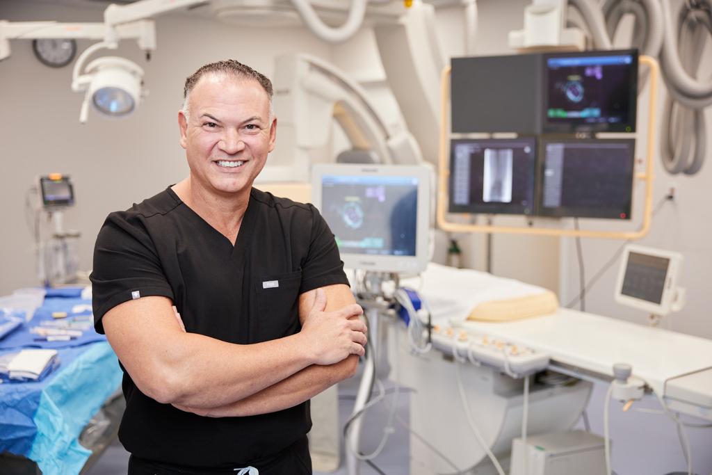
amavita Heart and Vascular Health® is a Miami interventional cardiology and vascular medicine practice led by Harvard-trained Dr. Pedro Martinez-Clark, MD, FACC, with two Miami-Dade locations in Kendall and North Miami Beach. Call (305) 290-4959.
Dr. Pedro Martinez-Clark is a Harvard-trained, board-certified interventional cardiologist in Miami, FL with over 15 years of experience.
Our Expertise
Expertise That Changes Lives
Where medical excellence meets genuine compassion
Led by Harvard-trained Dr. Pedro Martinez-Clark, a board-certified interventional cardiologist and Fellow of the American College of Cardiology (FACC), our team combines advanced techniques with personalized care to deliver exceptional outcomes.
About Dr. Martinez-Clark
Board-certified interventional cardiologist with over 15 years of experience. Harvard-trained at Massachusetts General Hospital. Fellow of the American College of Cardiology (FACC). Specializes in minimally invasive cardiac catheterization, coronary revascularization, peripheral vascular interventions, and structural heart disease. Works alongside Dr. William O'Neill, a world-renowned pioneer in interventional cardiology. Serves patients across Miami-Dade County from three convenient locations.
Minimally Invasive
Procedures with faster recovery times
Same-Day Outpatient
Greater comfort and convenience
15+ Years Experience
Complex cardiovascular care expertise
Multilingual Care
English, Spanish, and Haitian Creole
Our Physicians
Meet Our Doctors
World-class interventional cardiologists combining decades of pioneering research with compassionate, personalized patient care.
Harvard Medical School-trained interventional cardiologist with 27+ years of clinical excellence
ABIM Board-Certified in Internal Medicine, Cardiovascular Disease, and Interventional Cardiology
Fellow of the American College of Cardiology (FACC)
Affiliated with Mercy Hospital, Miami (HCA Florida Healthcare)
12,000+ minimally invasive cardiovascular procedures performed
Founder & CEO of the amavita CardioElite™ program for skilled nursing facilities
Leading Principal Investigator in cardiovascular clinical trials and frequent ACC presenter
Multilingual care in English, Spanish, and Haitian Creole
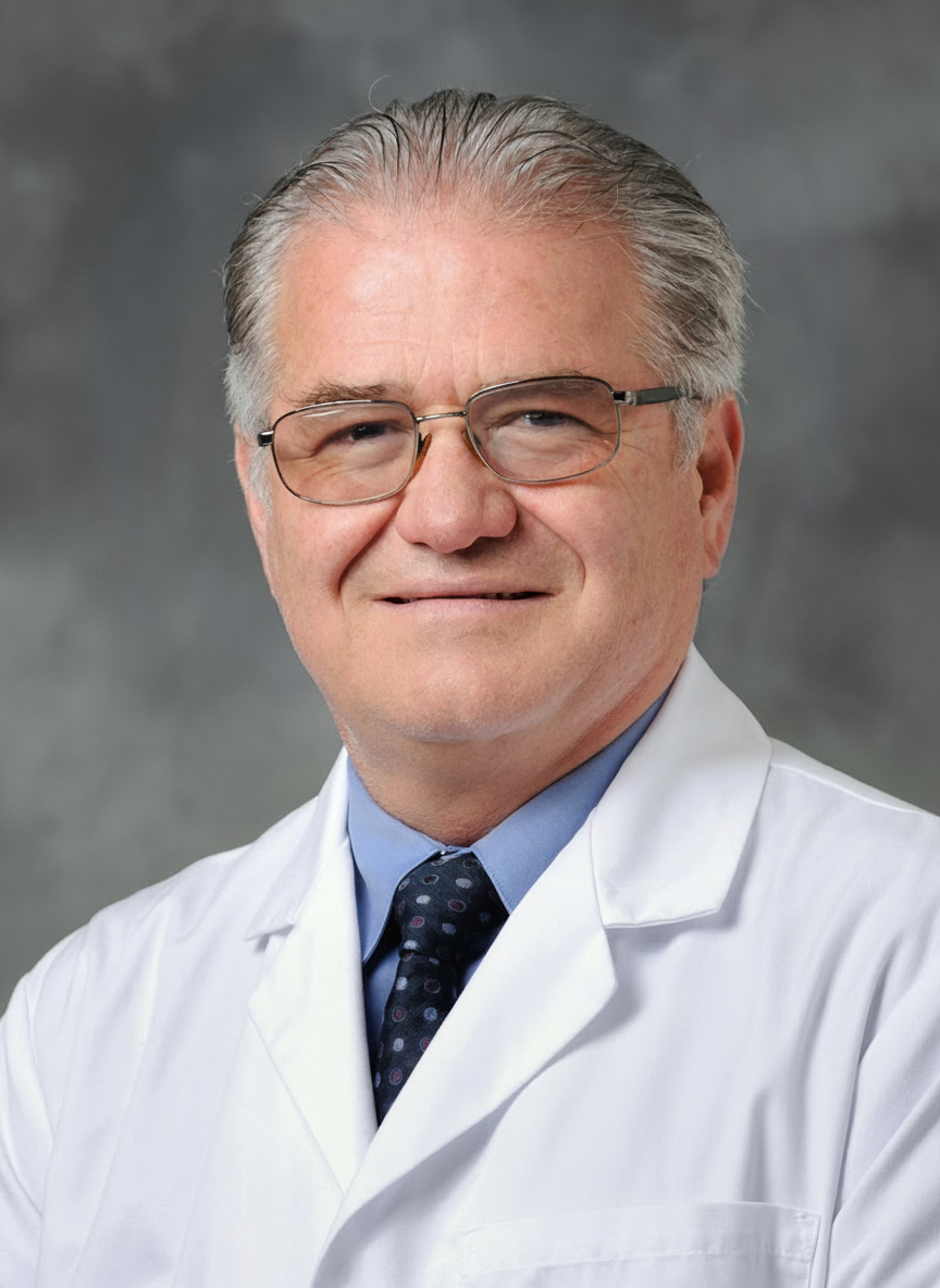
Medical Director, Center for Structural Heart Disease, Henry Ford Hospital (2012–present)
Pioneered angioplasty for heart attacks — now the worldwide standard of care
Performed the first transcatheter aortic valve replacement (TAVR) in the United States (2005)
National Principal Investigator for the Edwards PARTNER, Protect II, and Protect IV trials
Former Executive Dean for Clinical Affairs, University of Miami Miller School of Medicine
Recipient of the TCT Lifetime Achievement Award; author of 300+ peer-reviewed publications
Master Fellow of SCAI (MSCAI) with 45+ years of cardiovascular medicine experience
Chief Medical Officer of the amavita CardioElite™ program
Insurance Accepted
We Accept Most Major Insurance Plans
Our team will verify your coverage and discuss any costs before your visit. (305) 290-4959 to confirm your plan.
What We Offer
Comprehensive Cardiovascular Solutions
From advanced heart procedures to cutting-edge vascular treatments, our Miami cardiology practice offers a full spectrum of cardiovascular care under one roof.
Heart Care
Experience improved energy, mobility, and peace of mind.
Advanced treatment for coronary artery disease, valve and structural heart disease, and arrhythmias using minimally invasive procedures—all designed to improve heart function without major surgery.
Explore Heart ServicesVascular Care
Feel less pain, regain confidence, prevent serious complications.
Specialized diagnosis and treatment of peripheral artery disease and chronic venous insufficiency through advanced procedures to improve circulation and relieve symptoms.
Explore Vascular ServicesResearch
Access tomorrow's treatments today.
Access innovative treatments through our active clinical trials. Our team works with sponsors and patients to discover new concepts that advance cardiovascular care.
Explore ResearchLongevity
Live longer, live better.
Our longevity program focuses on proactive cardiovascular health, combining advanced diagnostics with lifestyle optimization to help you live a longer, more vibrant life.
Explore LongevityCardioElite™ for SNFs
AI-powered cardiac care for skilled nursing facilities.
Our amavita CardioElite™ program brings FDA-cleared AI cardiac diagnostics, 24/7 cardiologist support, and heart failure certification guidance to skilled nursing facilities across Florida — reducing readmissions by up to 70% at zero cost to partner facilities.
Visit CardioElite SiteGenicular Artery Embolization
Minimally invasive knee pain relief.
An innovative, non-surgical treatment for chronic knee pain caused by osteoarthritis. This catheter-based procedure reduces inflammation and pain without surgery or joint replacement.
Learn MoreSpecializations
Conditions We Treat
As a leading cardiologist in Miami, Dr. Martinez-Clark and the amavita HEART AND VASCULAR HEALTH® team treat the full range of heart and vascular conditions using the latest minimally invasive techniques.
"I drove all the way from Fort Myers because it was worth it."
— Gerardo Fedon, verified patient
Patient Stories
What Our Patients Say
"Dr. Pedro Martinez-Clark and the team at amavita are exceptional! From the moment you walk in, you're greeted with professionalism and warmth. Dr. Martinez-Clark is incredibly knowledgeable, attentive, and genuinely cares about his patients' well-being."
Jhon Tovar
Cardiac Catheterization Patient
"Courteous, friendly, and professional staff. Bright and clean office. Had several test procedures at this location with no issues. Everything went smoothly. I drove all the way from Fort Myers because it was worth it."
Gerardo Fedon
Cardiac Stress Testing Patient
"When I met Dr. Martinez Clark I was in such pain. After my first visit he assured me he would be able to help me feel better. After my procedure I am able to walk. I am thankful for his kindness and support. His staff is amazing."
Angela Zigler
Vascular Care Patient
Insights
Expert Cardiovascular Insights
Evidence-based articles on heart health, prevention, and living well
4 Key Differences Between Sudden Cardiac Arrest vs Heart Attack
Understanding the critical differences between cardiac arrest and heart attack can save lives. Learn the warning signs and when to seek emergency care.
Read MoreUnderstanding the Signs and Symptoms of AFib
Atrial fibrillation affects millions of Americans. Discover the common symptoms, risk factors, and modern treatment options available.
Read MoreUnderstanding GAE for Knee Pain Relief: A Complete Guide
Genicular artery embolization offers a minimally invasive alternative to knee surgery. Learn how this innovative procedure works.
Read MoreWhy Choose Us
The amavita Heart and Vascular Health® Difference
At amavita HEART AND VASCULAR HEALTH®—which means "love life" in Latin—we believe cardiovascular care should not only extend life but enhance its quality. We bring hospital-quality care into a more accessible, comfortable outpatient setting where you're treated as a person, not just a patient.
Patient-Centered
Decision making that respects your preferences and goals
Honest Communication
Delivered with compassion and hope
Technology + Care
Integration for the best possible outcomes
Community Commitment
Dedicated to improving cardiovascular health across Miami-Dade
Find Us
Our Locations
Conveniently located across Miami-Dade County — serving Kendall and North Miami Beach.
NorthPark Professional
100 NW 170th Street, Suite 305
North Miami Beach, FL 33169
Our Facility
State-of-the-Art Surgery Center
Miami's Premier Cardiovascular and Radiology Surgery Center

Advanced Cardiovascular of Miami
Miami's Premier Cardiovascular and Radiology Surgery Center
A next-generation cardiovascular and interventional radiology lab powered by expert physicians, exceptional staff, and state-of-the-art equipment. Our ambulatory surgery center brings hospital-quality procedures into a comfortable, efficient outpatient setting.
Co-located at our Kendall office — 9408 SW 87th Ave, Suite 303, Miami, FL 33176
Cardiology
- Cardiac Catheterization
- Cardiac Mapping & Ablation
- Coronary Intervention
- Implantable Cardiac Monitors
- Pacemakers/ICDs/Bi-Ventricular Devices
- Peripheral Artery & Vein Angiography
- Peripheral Artery & Vein Intervention
Interventional Radiology
- Genicular Artery Embolization
- Prostate Artery Embolization
- Uterine Fibroid Embolization
- Hemorrhoidal Artery Embolization
- Kyphoplasty
- Spinal Cord Stimulator Implantation
- Dialysis Access Management
Take the First Step Towards the Life You Want
Every day you wait could make a difference. Schedule your consultation today.
Whether you're experiencing symptoms, seeking a second opinion, or looking for preventative care, Dr. Martinez-Clark and the amavita HEART AND VASCULAR HEALTH® team are ready to provide you with world-class cardiovascular care.
Or call us directly at (305) 290-4959
Common Questions