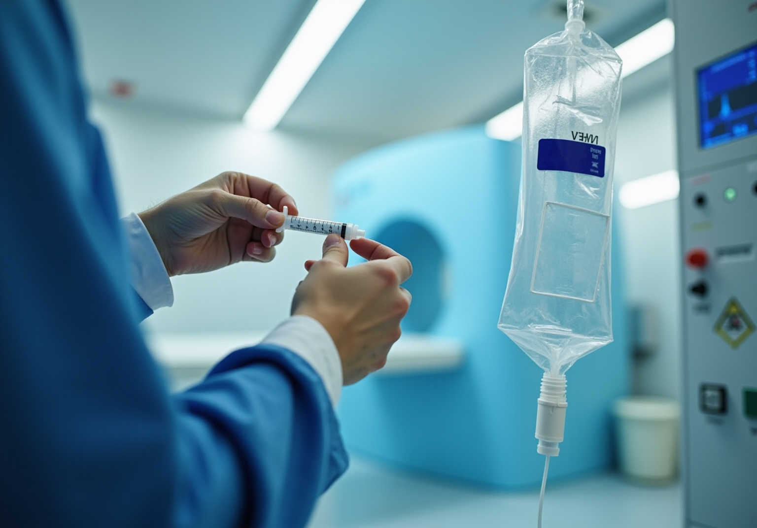


A PET heart scan is a gentle, non-invasive imaging test designed to assess heart function and blood circulation. This is crucial for diagnosing cardiovascular issues such as coronary artery disease and heart failure, which can understandably cause concern. This article highlights the importance of PET scans by illustrating how they provide comprehensive insights into cardiac health, enhance diagnostic accuracy, and enable personalized treatment plans. Ultimately, these benefits lead to improved patient outcomes, fostering a sense of hope and reassurance.
Have you ever felt anxious about your heart health? It’s completely normal to have such feelings. Understanding your heart's condition can be daunting, but PET scans offer a clear view of what’s happening inside your body. By using this advanced technology, healthcare professionals can better understand your unique situation and tailor the best approach for your care.
Furthermore, knowing that you have access to such detailed information can empower you to take charge of your health. With the insights gained from a PET scan, doctors can create a treatment plan that’s just right for you, ensuring that you receive the care you truly deserve. Remember, you are not alone in this journey; there are compassionate professionals ready to support you every step of the way.
A Positron Emission Tomography (PET) heart scan is an important advancement in cardiovascular diagnostics. This non-invasive method allows us to visualize heart function and blood flow, which can be a source of concern for many patients. By enhancing our understanding of various cardiac conditions, this innovative imaging technique empowers healthcare providers to create personalized treatment plans that can lead to improved patient outcomes.
However, as we increasingly rely on such technology, it’s essential to consider its implications. What challenges and opportunities does this present for both patients and practitioners in the evolving landscape of heart health? We are here to support you through these changes, ensuring that you feel informed and cared for every step of the way.
A Positron Emission Tomography (PET) heart scan is a non-invasive imaging test designed to assess your heart's function and blood circulation. This advanced method involves a minor injection of a radioactive substance into your bloodstream, allowing for dynamic images of your heart to be captured. These images can reveal areas with reduced blood flow or unusual metabolic activity, providing essential insights into your body's physiological processes.
Unlike traditional imaging techniques, PET examinations are particularly effective in , such as coronary artery disease and heart failure. They offer a more comprehensive view of your cardiac health, which can be reassuring during times of uncertainty.
In the United States, the use of cardiac PET imaging has significantly increased, with about a 25% growth from 2018 to 2022. This rise reflects the growing importance of these examinations in clinical practice. Cardiologists have found that PET imaging can greatly enhance diagnostic accuracy, especially when standard evaluations raise concerns. For instance, a typical PET examination reveals uniformly distributed blood flow to the heart muscle, while irregular results may suggest blocked or narrowed arteries, prompting further assessment and management.
Recent advancements in PET technology, such as integrated PET-CT systems, allow for simultaneous evaluation of both anatomical and functional aspects of coronary artery disease. This improvement aids in risk stratification and treatment planning, making the PET heart scan an invaluable resource in modern cardiology. It empowers healthcare professionals to make informed decisions that can ultimately enhance your health outcomes.
Before the procedure, you may be advised to limit caffeine intake for 24 hours. Additionally, it’s recommended to avoid close contact with infants or expectant mothers for a few hours afterward. Most individuals can resume their normal activities immediately after the examination. However, if a sedative is used, it’s important to avoid driving, operating heavy machinery, and consuming alcohol for at least 24 hours. After your examination, consider scheduling a follow-up appointment with your physician to discuss the findings and any necessary next steps. Remember, you are not alone in this journey, and support is always available.
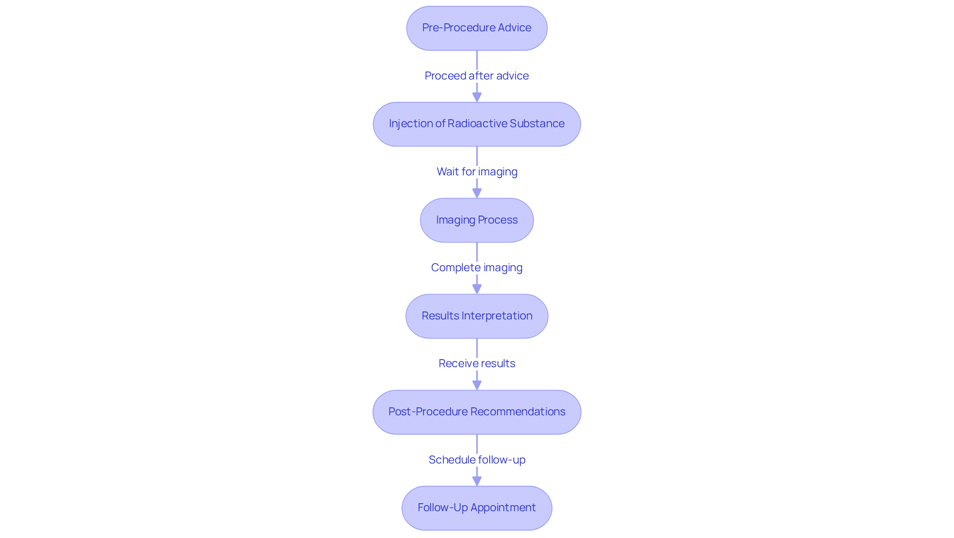
The pet heart scan has become an essential resource in promoting cardiovascular well-being, especially as our aging population faces an increasing incidence of cardiac illness. These examinations provide a comprehensive understanding of heart performance, which is crucial for older individuals who often experience various comorbidities that complicate traditional diagnostic approaches. By offering a clearer and more detailed view of cardiac health, PET scans empower healthcare providers to make informed treatment decisions, ultimately leading to improved health outcomes. This capability is particularly significant, as advanced diagnostic tools can facilitate more tailored treatment plans and enhance the management of cardiovascular conditions.
Amavita's CardioElite™ program further enhances this process by integrating advanced imaging capabilities with 24/7 cardiology consultation. This initiative serves as a clinical force multiplier, ensuring that high-risk individuals receive thorough evaluations and . For instance, research indicates that combining coronary artery calcium (CAC) scoring with myocardial perfusion imaging can significantly improve risk assessment, especially in individuals with normal perfusion results but elevated CAC scores. Such advancements enable early identification of potential issues and support clinical decision-making, ultimately enhancing the quality of care for elderly individuals.
Additionally, cardiac PET boasts an average sensitivity of 89% and specificity of 90% for detecting obstructive coronary artery disease, highlighting its effectiveness in improving patient outcomes. As the field of cardiovascular diagnostics progresses, the role of PET imaging continues to expand, underscoring its importance in enhancing health results for the elderly, particularly when supported by comprehensive initiatives like CardioElite™. Remember, you are not alone in this journey; we are here to support you every step of the way.
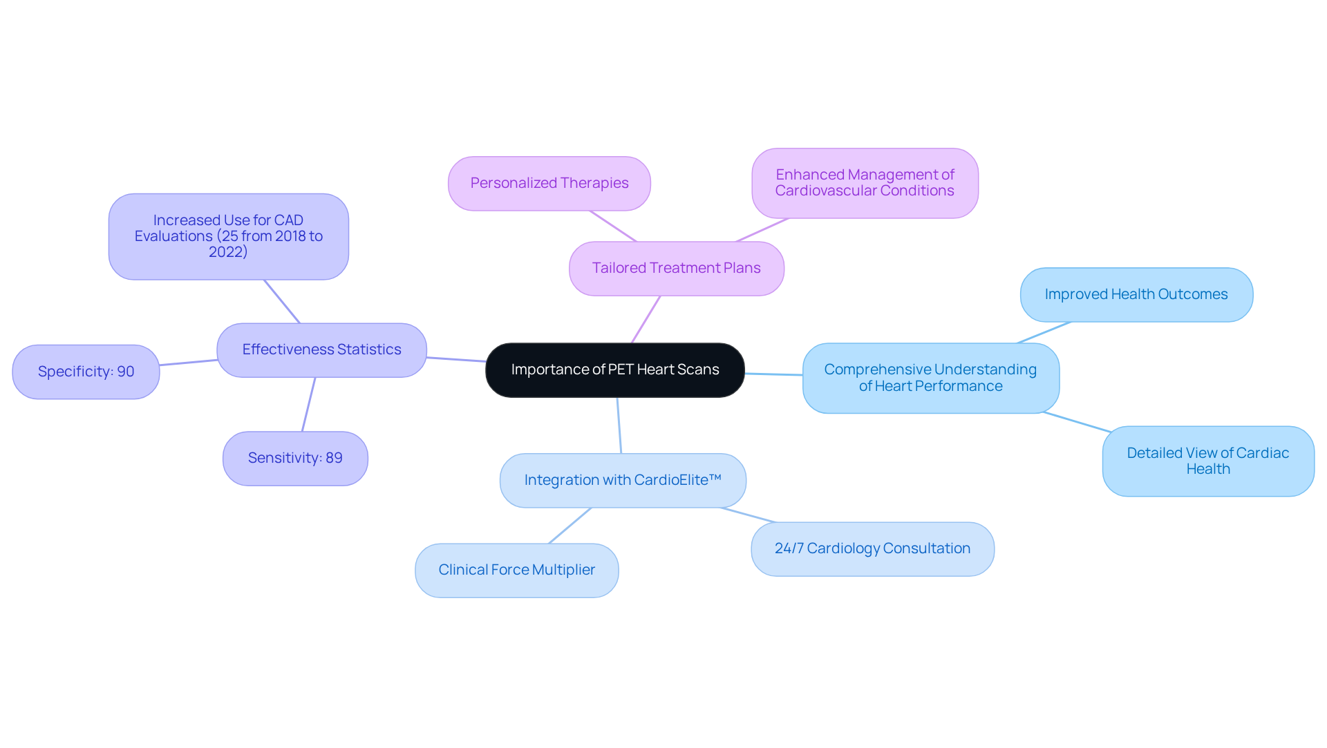
The benefits of PET cardiac imaging are wide-ranging and substantial, offering precise insights into blood circulation and cardiac function that are crucial for diagnosing conditions such as coronary artery disease (CAD). At Amavita Heart and Vascular Health®, we understand how important it is to detect regions of the heart at risk of harm. Our cardiac PET imaging has been vital in enabling prompt actions that can avert additional complications. Our specialists employ advanced diagnostic imaging methods to ensure that you receive the most effective care possible, all while ensuring minimal discomfort during these non-invasive examinations, allowing you to resume your daily activities shortly after the procedure.
In addition to this, PET imaging plays a crucial role in guiding treatment decisions. By providing detailed information about your heart's condition, we can determine whether surgical interventions, such as stenting or bypass surgery, are necessary or if medical therapies would suffice. This tailored approach not only enhances treatment outcomes but also contributes to a more efficient healthcare experience, reducing the need for invasive procedures. Overall, the incorporation of a PET heart scan into healthcare illustrates our dedication to enhancing your cardiovascular well-being through advanced diagnostic methods.
However, it is important to consider the potential risks associated with PET imaging, such as heartbeat disorders and the impact of recent food intake on results. Additionally, the procedure typically takes between 30 minutes to an hour, with results available in less than 45 minutes—significantly faster than traditional imaging techniques that can take 3-4 hours. This efficiency, along with the capacity to address various healthcare demographics, including individuals with obesity or intricate coronary artery conditions, highlights the significance of PET imaging in contemporary cardiovascular treatment. We are here to , ensuring that your health is prioritized.
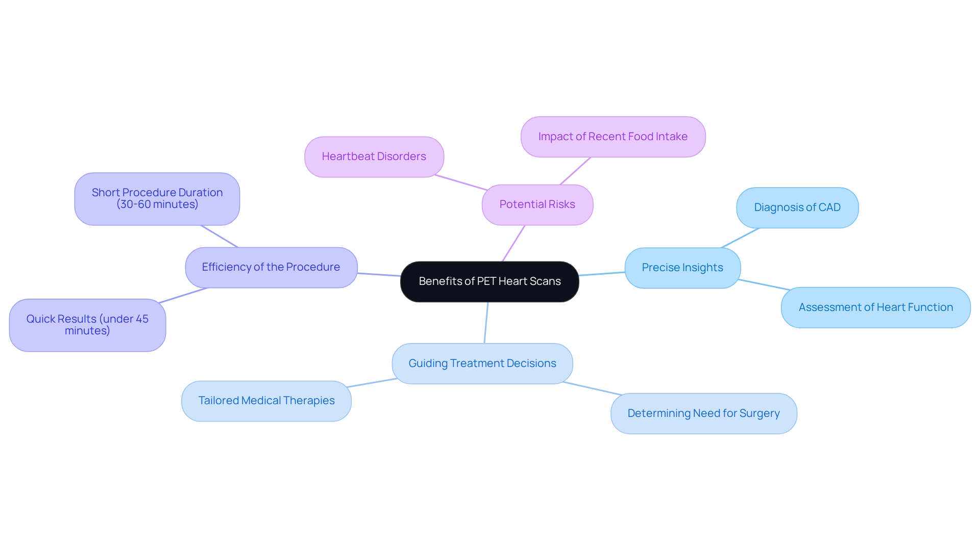
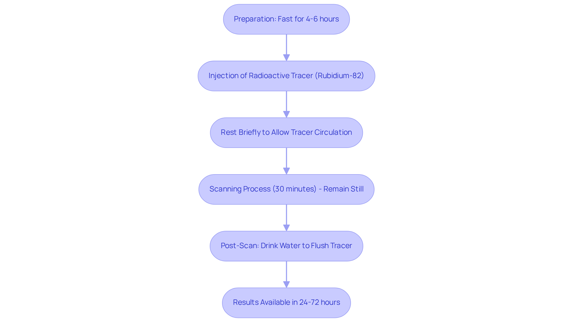
A pet heart scan is designed to be a straightforward and user-friendly process, particularly beneficial for individuals at high risk due to conditions like diabetes, hypertension, or a family history of heart disease. Initially, a small amount of radioactive tracer, such as Rubidium-82, is gently injected into a vein, which typically takes just a few minutes. After the injection, individuals are encouraged to rest briefly, allowing the tracer to circulate effectively throughout their bodies. The actual scanning process involves lying on a table that moves through a large, donut-shaped machine known as a PET scanner. This scanning phase usually lasts around 30 minutes, during which it is essential for individuals to remain still to ensure the captured.
The entire PET heart examination procedure typically requires under one hour. Once the scan is completed, individuals can promptly return to their regular activities, as the amount of radioactive material used is minimal and poses little risk. Most people experience no adverse effects from the tracer, although the IV insertion site may feel slightly sore or bruised. Healthcare professionals will discuss the results with individuals, which are usually accessible within 24 to 72 hours, helping them understand the implications for their cardiovascular health and guiding any necessary follow-up measures.
Patients are advised to fast for 4 to 6 hours prior to the examination to enhance the accuracy of the results. Additionally, after the PET imaging procedure, individuals are encouraged to drink several glasses of water to assist in flushing out the residual radioactive tracer from their bodies.
The pet heart scan not only assists in diagnosing conditions such as coronary artery disease but also offers valuable insights into heart function, enabling personalized treatment plans. This non-invasive imaging technique, such as the pet heart scan, is recognized for its high diagnostic accuracy and low radiation exposure, making it a preferred choice for assessing cardiovascular health. At Amavita Heart and Vascular Health®, we integrate advanced imaging capabilities into our comprehensive care plans, ensuring that each patient receives personalized attention and innovative treatment options. This approach is especially beneficial for those seeking executive health screenings or monitoring treatment effectiveness.
A PET heart scan is an invaluable tool for assessing cardiovascular health, offering a non-invasive way to visualize heart function and blood flow. This advanced imaging technique not only enhances diagnostic accuracy but also empowers healthcare professionals to make informed decisions about your care. By providing a clearer picture of cardiac conditions, PET scans play a crucial role in managing heart disease and improving overall health outcomes.
Throughout our discussion, we have highlighted the importance of PET heart scans in diagnosing conditions such as coronary artery disease and heart failure. The efficiency of this procedure, along with its ability to deliver detailed information about your cardiac health, underscores its growing significance in modern cardiology. Furthermore, advancements in technology, such as integrated PET-CT systems, enhance the diagnostic capabilities of PET imaging, allowing for comprehensive evaluations that guide treatment decisions.
As cardiovascular diseases become more prevalent, embracing innovative diagnostic methods like PET heart scans is essential. These scans not only provide critical insights into your heart function but also facilitate timely interventions that can prevent complications. If you are at risk or seeking to understand your cardiac health better, we encourage you to engage with your healthcare provider about the benefits of PET heart scans. This can lead to more personalized and effective care strategies. By prioritizing your heart health through advanced imaging, you can pave the way for improved quality of life and longevity.
What is a PET heart scan?
A PET heart scan is a non-invasive imaging test that assesses the heart's function and blood circulation using a minor injection of a radioactive substance to capture dynamic images of the heart.
How does a PET heart scan differ from traditional imaging techniques?
Unlike traditional imaging methods, PET scans are particularly effective in diagnosing cardiovascular issues such as coronary artery disease and heart failure, providing a more comprehensive view of cardiac health.
What are the benefits of using PET imaging in cardiology?
PET imaging enhances diagnostic accuracy, especially when standard evaluations raise concerns. It can reveal blood flow distribution to the heart muscle and indicate potential blockages or narrowed arteries.
How has the use of cardiac PET imaging changed in recent years?
In the United States, the use of cardiac PET imaging has increased significantly, with about a 25% growth from 2018 to 2022, reflecting its growing importance in clinical practice.
What advancements have been made in PET technology?
Recent advancements include integrated PET-CT systems that allow for simultaneous evaluation of both anatomical and functional aspects of coronary artery disease, aiding in risk stratification and treatment planning.
What preparations are needed before a PET heart scan?
Patients may be advised to limit caffeine intake for 24 hours before the procedure and to avoid close contact with infants or expectant mothers for a few hours afterward.
What should I expect after a PET heart scan?
Most individuals can resume normal activities immediately after the examination. However, if a sedative was used, it is important to avoid driving, operating heavy machinery, and consuming alcohol for at least 24 hours.
Should I follow up with my physician after the PET heart scan?
Yes, it is recommended to schedule a follow-up appointment with your physician to discuss the findings of the scan and any necessary next steps.