

PET scans are essential in cardiology, playing a vital role in understanding blood circulation and evaluating the health of the heart. They help in diagnosing coronary artery disease with remarkable accuracy, offering peace of mind to patients and their families. With the advancement of tracers and non-invasive procedures, these scans become even more effective, especially for our elderly loved ones. This means that patients can receive care that is not only precise but also tailored to their individual needs, based on detailed functional imaging.
In addition to this, the ability to assess myocardial viability ensures that treatment plans can be customized, providing reassurance that every patient is receiving the best possible care. This personalized approach makes a significant difference in the quality of life for those who may feel anxious about their health. Remember, it’s perfectly normal to have concerns, and reaching out for support can be a vital step towards better health. Together, we can navigate these challenges with compassion and understanding.
The landscape of cardiac diagnostics has been transformed by the introduction of Positron Emission Tomography (PET) scans, which provide invaluable insights into heart health. These advanced imaging techniques not only assess blood flow and myocardial viability but also play a vital role in accurately diagnosing coronary artery disease.
As you consider your heart health, you may have questions:
This guide aims to address these concerns, highlighting the importance of PET scans in cardiology, outlining the procedural steps, and exploring the many benefits they offer for proactive cardiac care. We are here to support you every step of the way.
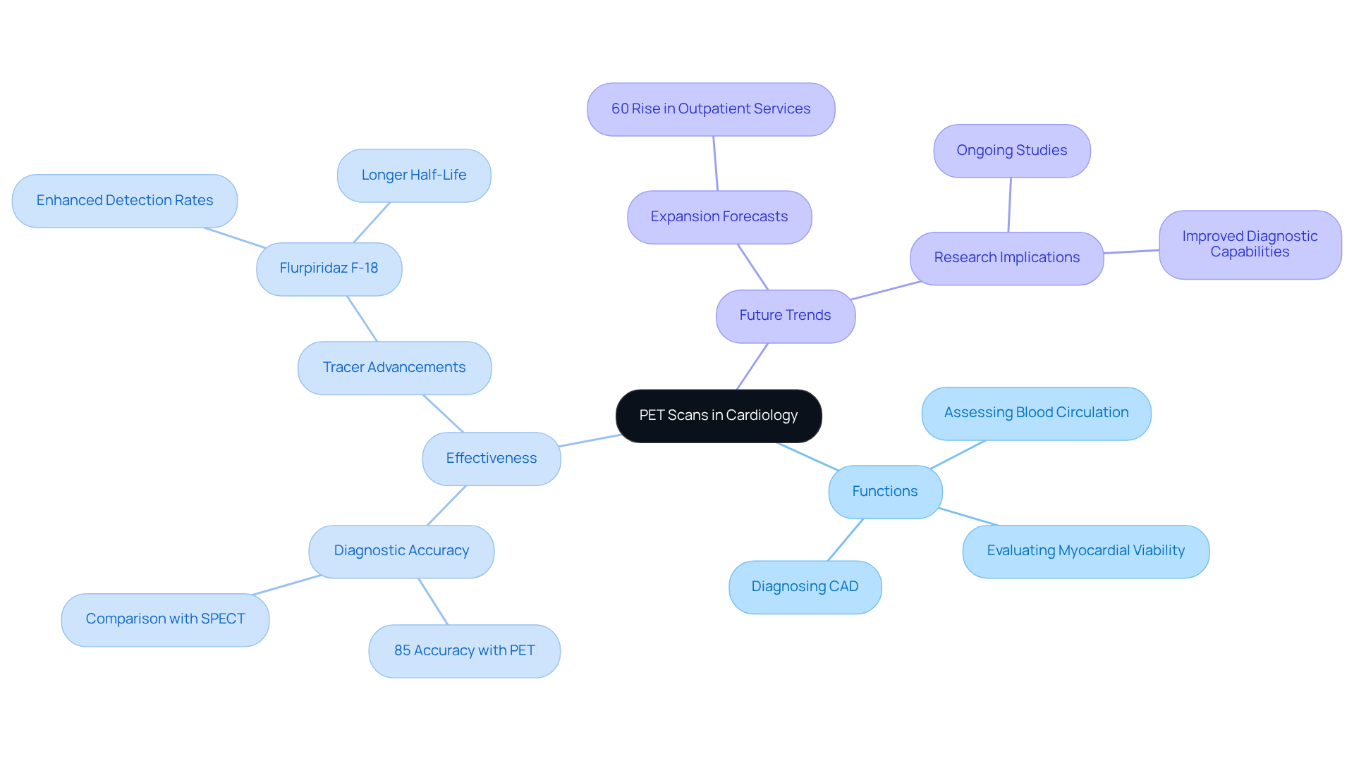
Positron Emission Tomography (PET) imaging represents a significant advancement in cardiac visualization, employing radioactive tracers to illustrate metabolic processes within the body. In cardiology, PET scan cardiology is essential for:
By providing high-resolution images that illustrate both the functional and structural features of the heart, PET scan cardiology examinations enable healthcare professionals to make informed treatment choices, especially for individuals with intricate cardiovascular conditions. This non-invasive procedure is particularly beneficial for elderly populations, where traditional diagnostic methods may pose greater risks or yield less accurate results.
Have you ever felt anxious about your heart health? Recent studies emphasize the effectiveness of PET scan cardiology in detecting CAD, demonstrating a diagnostic accuracy of 85%, significantly surpassing that of traditional methods like single-photon emission computed tomography (SPECT). For example, the introduction of the new tracer flurpiridaz F-18 has shown enhanced detection rates in PET scan cardiology, particularly in women and individuals with obesity, thereby improving overall diagnostic capabilities.
Real-world applications of PET scans have proven transformative. The PET scan cardiology is recommended for individuals experiencing symptoms such as chest pain or shortness of breath, as it helps identify possible blockages in the heart arteries. Furthermore, this advanced imaging technology, such as PET scan cardiology, enables earlier and more precise detection of CAD, allowing for customized treatment plans that align with individual requirements. As the use of PET scan cardiology continues to expand, with forecasts suggesting a 60% rise in outpatient services over the next five years, ongoing research is anticipated to further enhance its applications in evaluating coronary artery disease and improving outcomes for individuals. Remember, you are not alone in this journey, and there are compassionate professionals ready to support you every step of the way.

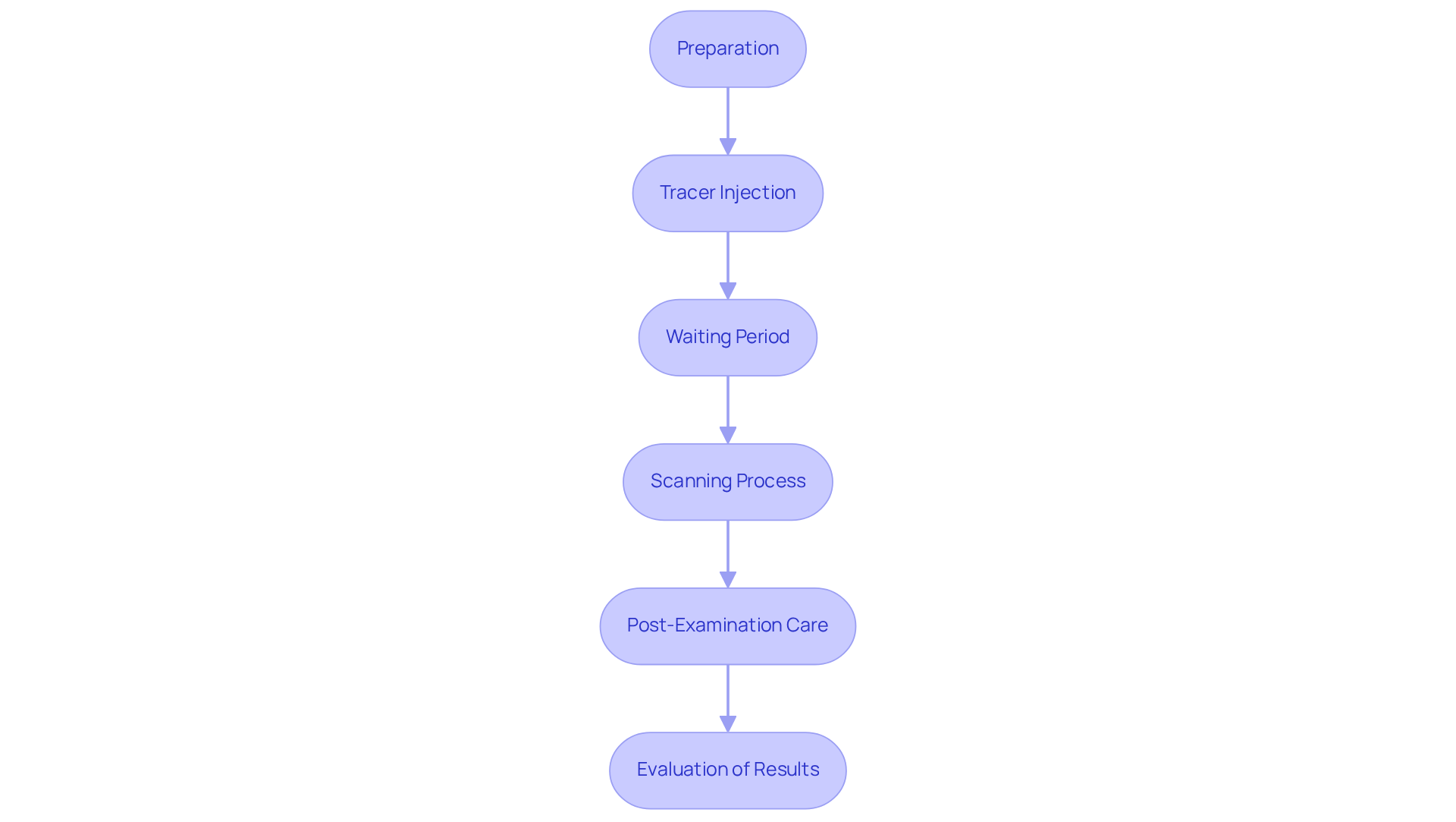
The procedure for pet scan cardiology involves several essential steps designed to ensure accurate results and your comfort, particularly if you are a high-risk patient, such as those with diabetes, hypertension, or a family history of heart disease.
First, preparation is key. Patients are generally recommended to fast for several hours before the examination and to avoid intense activities. This preparation helps ensure that the radioactive tracer used during the scan provides precise imaging results.
Next comes the tracer injection. A small amount of radioactive tracer is injected into a vein, usually in the arm. This tracer emits positrons, which are detected by the PET scan cardiology system, enabling detailed imaging of the heart. Although allergic responses to the tracer are uncommon, it’s important to notify your healthcare provider of any previous allergies.
After the injection, there is a waiting period. Patients usually wait for about 30 to 60 minutes. This waiting duration enables the tracer to circulate and accumulate in the cardiac tissue, thereby improving the quality of the images obtained during the pet scan cardiology examination.
Then, the scanning process begins. Patients lie on a table that slides into the pet scan cardiology device. This pet scan cardiology process takes approximately 30 to 60 minutes, during which the machine captures images of the heart, highlighting areas of blood flow as the tracer accumulates in those regions. In certain situations, pet scan cardiology may be combined with MRI or CT examinations to enhance image analysis.
Finally, after the examination, you can typically return to your usual activities. It is important to stay hydrated after the procedure, as this helps flush the tracer from your body. A cardiologist will examine the images obtained through pet scan cardiology to evaluate the condition of your heart and decide on any required follow-up actions.
You can feel reassured that the PET scan is a painless and , with minimal side effects associated with the tracer. The procedure generally entails low radiation exposure, making it a safe choice for evaluating cardiovascular health using pet scan cardiology. This experience is designed to be as comfortable as possible, allowing for effective assessment of heart health, especially for those seeking comprehensive cardiovascular care at Amavita.

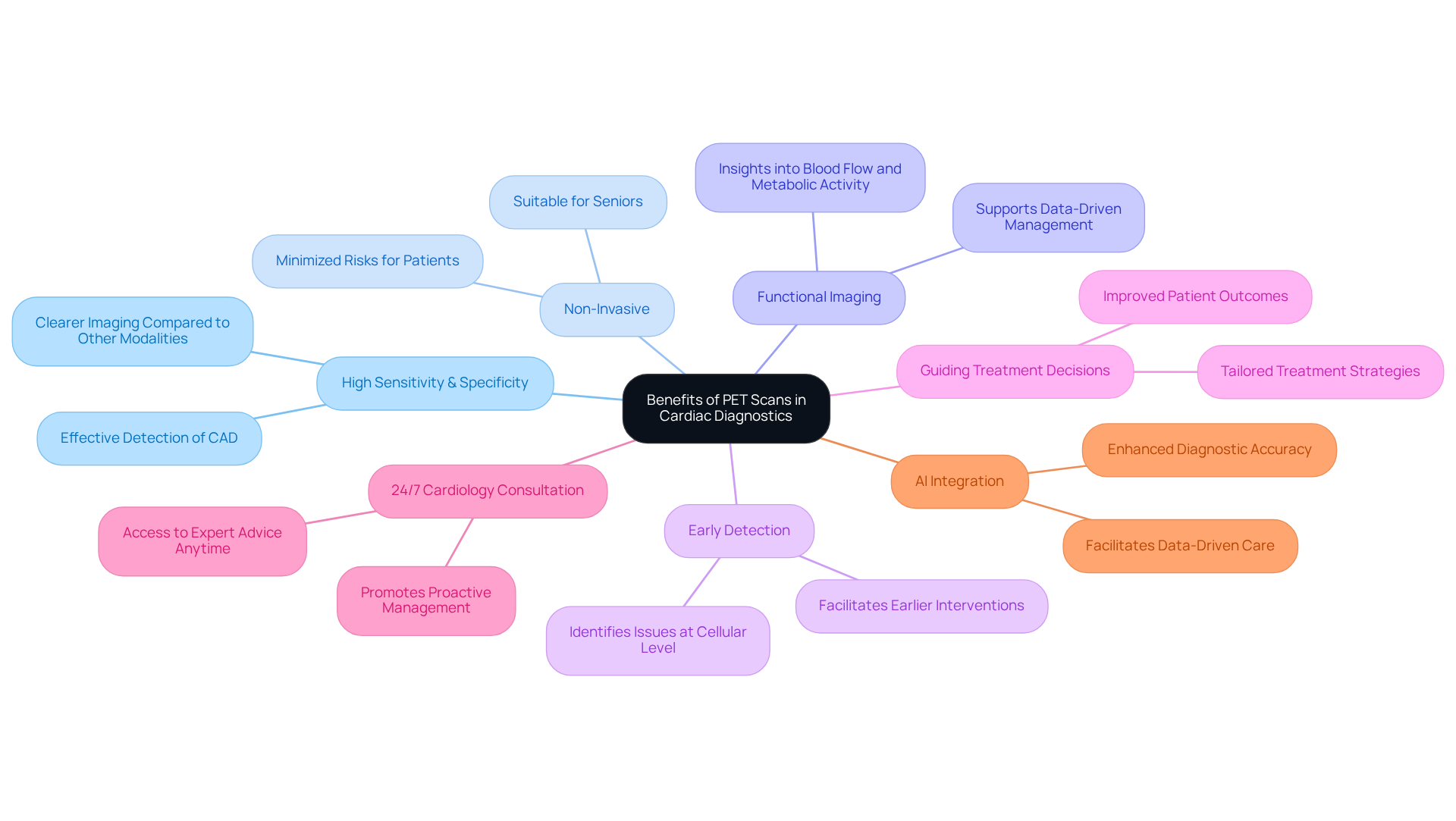
The benefits of pet scan cardiology are significant in cardiac diagnostics, especially when integrated into comprehensive programs like Amavita's CardioElite™.
PET scan cardiology demonstrates high sensitivity and specificity, as PET scans are remarkably effective in detecting coronary artery disease and assessing myocardial perfusion, offering clearer images than many other imaging modalities. This accuracy is crucial for proactive management of cardiac individuals, helping to reduce the likelihood of readmissions.
In practical terms, PET imaging has shown effectiveness in diagnosing CAD, often serving as the gold standard for determining whether individuals should proceed with stenting or bypass surgery. As specialists in the field have pointed out, the integration of pet scan cardiology technology is transforming cardiac care, enhancing diagnostic accuracy and outcomes for individuals. This evolution underscores the growing recognition of pet scan cardiology as an essential tool in managing cardiovascular health.

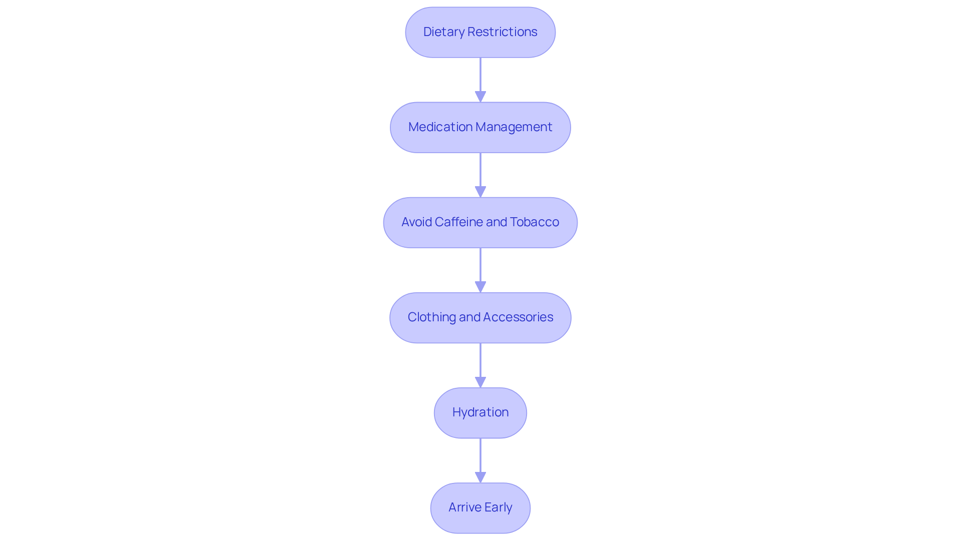
To ensure a successful PET scan, it’s important to follow these preparation guidelines with care:
By diligently following these guidelines, you can maximize the effectiveness and reliability of your PET scan cardiology results. Remember, we are here to support you every step of the way.

Understanding the role of PET scans in cardiology is essential for patients who seek clarity about their heart health. These advanced imaging techniques not only provide a detailed view of cardiac function but also enhance diagnostic accuracy, allowing healthcare providers to create personalized treatment plans. By utilizing radioactive tracers, PET scans unveil vital metabolic processes, playing a crucial role in assessing and managing coronary artery disease and other cardiovascular conditions.
Throughout this article, we have shared important insights regarding the effectiveness of PET scans in diagnosing heart issues, the procedural steps involved, and the significant benefits they offer compared to traditional methods. With an impressive diagnostic accuracy rate and the ability to detect conditions at an early stage, PET scans are truly transforming cardiac care. Moreover, the non-invasive nature of the procedure, coupled with comprehensive preparation guidelines, ensures patient safety and comfort, making it a preferred choice for many individuals, especially those at higher risk.
As the field of cardiology evolves, embracing innovative technologies like PET scans becomes increasingly vital. We encourage patients to engage with their healthcare professionals about their heart health and to consider PET scans as a valuable tool in proactive cardiac care. Understanding these advancements empowers individuals to make informed decisions and reinforces the importance of regular health assessments in safeguarding heart health. Remember, you are not alone on this journey; support is always available to help you navigate your heart health with confidence.
What is a PET scan and its role in cardiology?
A Positron Emission Tomography (PET) scan is an advanced imaging technique that uses radioactive tracers to visualize metabolic processes in the body. In cardiology, it is essential for assessing blood circulation to the heart muscle, evaluating myocardial viability, and diagnosing coronary artery disease (CAD).
How does PET scan cardiology benefit patients, especially the elderly?
PET scan cardiology provides high-resolution images that show both functional and structural features of the heart, aiding healthcare professionals in making informed treatment decisions. It is particularly beneficial for elderly patients, as traditional diagnostic methods may pose greater risks or yield less accurate results.
What is the diagnostic accuracy of PET scans in detecting coronary artery disease (CAD)?
Recent studies indicate that PET scan cardiology has a diagnostic accuracy of 85% for detecting CAD, which is significantly higher than traditional methods like single-photon emission computed tomography (SPECT).
How has the introduction of new tracers like flurpiridaz F-18 impacted PET scan cardiology?
The new tracer flurpiridaz F-18 has shown enhanced detection rates in PET scan cardiology, particularly benefiting women and individuals with obesity, thereby improving overall diagnostic capabilities.
When is the PET scan cardiology myocardial perfusion stress test recommended?
The PET scan cardiology myocardial perfusion stress test is recommended for individuals experiencing symptoms such as chest pain or shortness of breath, as it helps identify possible blockages in the heart arteries.
What are the future trends for PET scan cardiology?
The use of PET scan cardiology is expected to expand, with forecasts suggesting a 60% rise in outpatient services over the next five years. Ongoing research is anticipated to further enhance its applications in evaluating coronary artery disease and improving patient outcomes.