
Introduction
Echocardiograms play a crucial role in modern medicine, providing a gentle glimpse into the heart’s structure and function through non-invasive ultrasound technology. Among the different types, the bubble study is particularly noteworthy for its ability to enhance visualization of blood flow and identify potential heart defects, like patent foramen ovale (PFO). Yet, many individuals may feel uncertain about when these specialized tests are needed or how they are performed.
What insights can an echocardiogram with a bubble study reveal? Understanding this procedure can empower you on your journey to better cardiovascular health. It’s natural to have questions and concerns, and knowing what to expect can make a significant difference.
In addition to this, recognizing the importance of these tests can help you take proactive steps in managing your heart health. If you’re feeling anxious or hesitant, remember that you’re not alone. Many patients share similar feelings, and it’s perfectly okay to seek support.
Ultimately, understanding the role of echocardiograms and bubble studies can lead to better health outcomes. So, take a moment to reflect on your heart health and consider reaching out for guidance. Your well-being is important, and there are resources available to help you every step of the way.
Define Echocardiograms and Bubble Studies
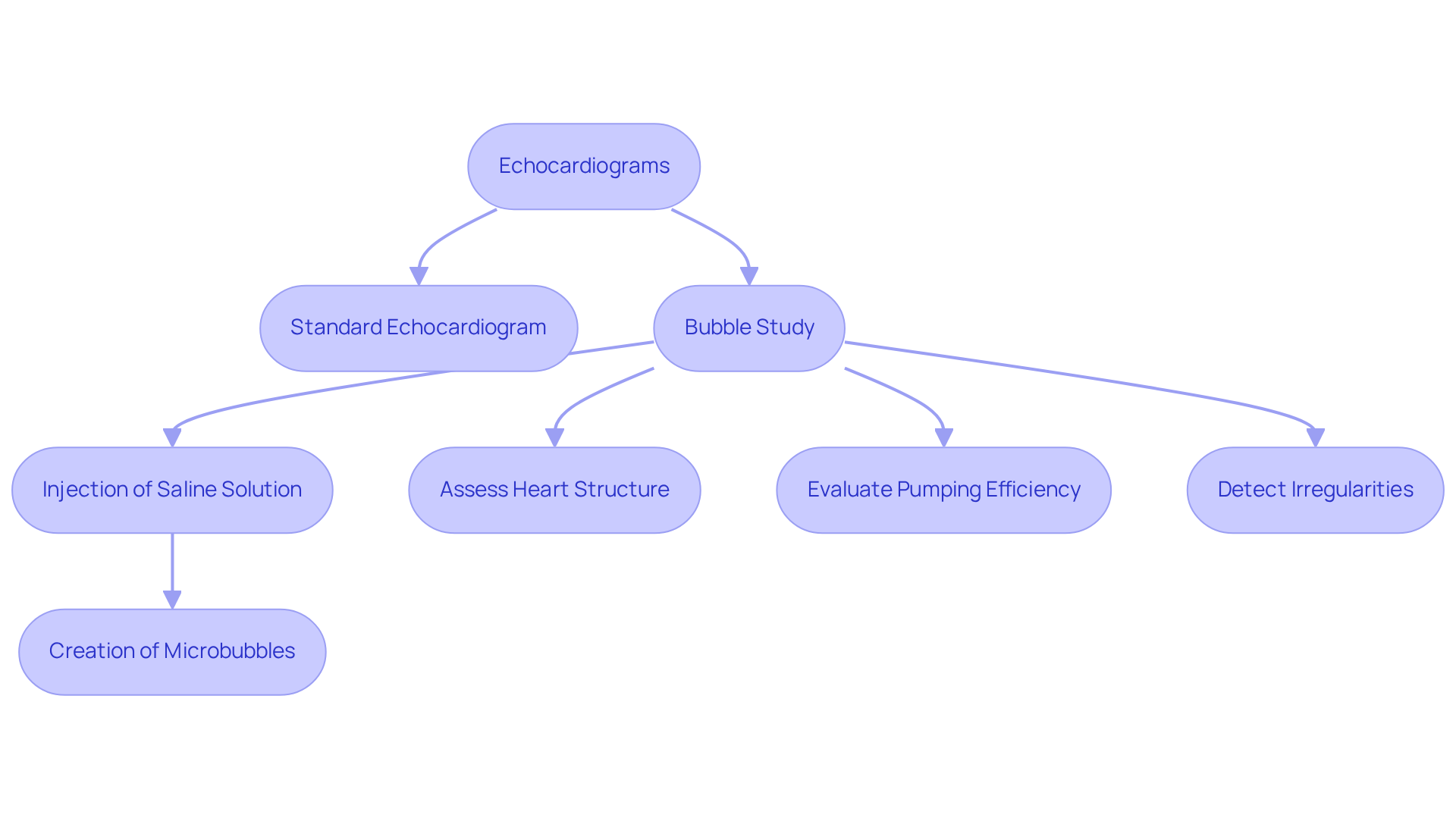
Echocardiograms are gentle, non-invasive ultrasound examinations that use high-frequency sound waves to create dynamic images of your heart. These tests are crucial for understanding how your heart is structured and how well it functions. They help us assess the size of the heart, the condition of the valves, and the overall pumping efficiency.
One specialized type of echocardiogram is where a saline solution mixed with air is injected into a vein. This creates tiny microbubbles that can be seen during the procedure, greatly enhancing our ability to visualize blood flow. This method is particularly helpful in detecting irregularities, such as shunts between the heart chambers. It’s especially effective for diagnosing conditions like patent foramen ovale (PFO) and other cardiac anomalies.
Recent data shows that bubble studies are performed in about 6% of echocardiographic examinations, highlighting their growing importance in clinical practice. For instance, in a study involving 663 individuals who experienced acute ischemic strokes, contrast examinations played a vital role in diagnosis, with 54 cases identified. This underscores their significance in managing stroke care.
At Amavita Heart and Vascular Health®, we understand that facing health issues can be daunting. That’s why we focus on comprehensive and preventive strategies tailored to your life. Our approach includes education and personalized interventions designed to support you every step of the way.
As research continues to evolve, the effectiveness of echocardiograms and contrast examinations in promoting cardiovascular health remains a priority. We are committed to advancing diagnostic methods and enhancing care for individuals like you. If you have any concerns about your heart health, please don’t hesitate to reach out. We’re here to help you navigate your health journey.
Identify Indications for Echo Bubble Studies
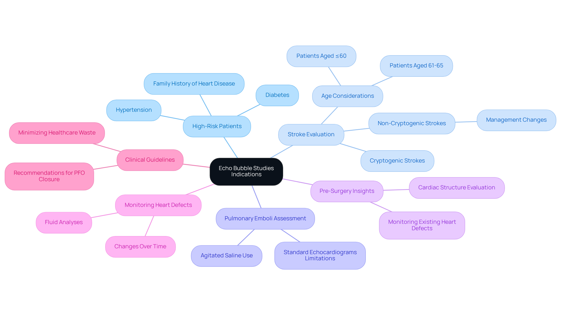
Echo assessments play a crucial role in many clinical situations, especially for those at higher risk, like individuals with diabetes, hypertension, or a family history of heart disease. These investigations, particularly the bubble study, are essential for identifying structural heart defects, such as patent foramen ovale (PFO) or atrial septal defects (ASD). If left undiagnosed, these conditions can lead to serious complications, and we want to ensure you’re aware of the importance of these evaluations.
Have you or a loved one experienced a stroke without any recognizable risk factors? If so, an echo bubble study can help reveal the presence of a shunt, thereby clarifying the cause of the stroke. Recent studies show that a significant number of patients didn’t change management. This highlights the need for further evaluation. Interestingly, only 32.8% of those who underwent PFO workup were aged 60 or younger, underscoring the importance of considering age in these evaluations.
These analyses are also beneficial in assessing potential abnormalities that standard echocardiograms might miss, providing a more comprehensive view of your condition. Additionally, fluid analyses can evaluate the heart’s pumping efficiency and identify any irregularities in blood circulation, which are vital for diagnosing various cardiovascular conditions.
For those preparing for surgery, contrast examinations offer essential insights into cardiac structure, ensuring that any existing issues are addressed before the procedure. If you have previously diagnosed heart conditions, echo contrast examinations are invaluable for monitoring changes over time, allowing for timely interventions.
If you’re experiencing unexplained symptoms like shortness of breath or fatigue, contrast examinations can provide important diagnostic insights that guide further management. Current guidelines recommend the use of an echo bubble study primarily for individuals with cryptogenic strokes, particularly those aged 18-60. It’s crucial to align clinical practice with established protocols to minimize unnecessary healthcare costs. In the U.S., healthcare spending on unnecessary tests is significant, with 20-30% considered waste. This emphasizes the need for thoughtful use of diagnostic resources.
As Jonathan Hu noted, there’s a considerable amount of diagnostic research that lacks backing from clinical guidelines. This further highlights the importance of carefully selecting individuals for these assessments. Remember, you’re not alone in navigating these health concerns, and seeking the right evaluations can make a significant difference in your care.
Explain the Procedure for Conducting an Echo Bubble Study
If you’re feeling anxious about the procedure, you’re not alone. Many patients share similar concerns, and it’s completely normal to have questions about what to expect. Let’s walk through the procedure together, step by step, so you can feel more at ease.
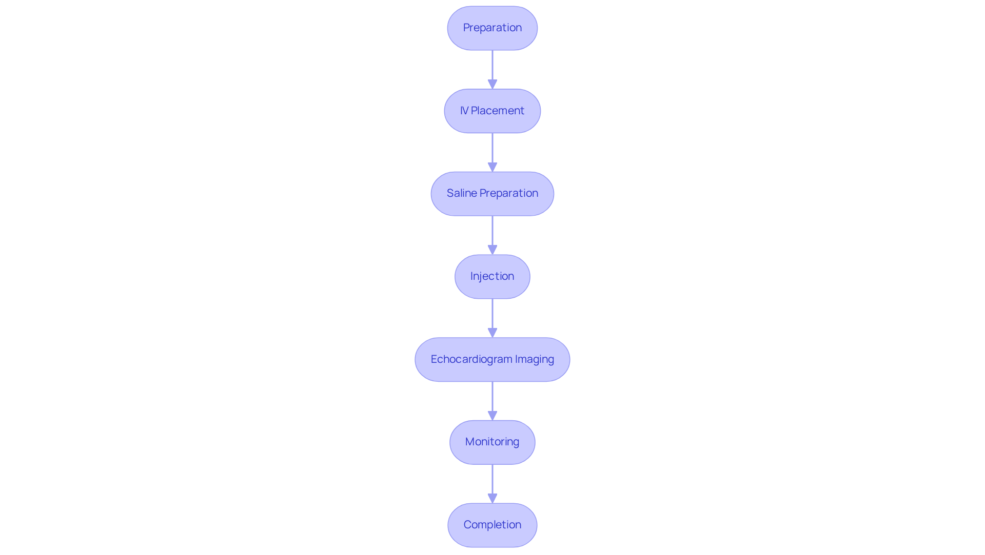
- Preparation: You’ll be comfortably positioned, usually lying on your left side. This position helps us get a clearer view of your heart, making the process smoother for you.
- Catheter Insertion: A small intravenous (IV) catheter will be gently inserted into a vein in your arm. This is a quick step, and we’ll ensure you’re as comfortable as possible.
- Saline Preparation: We prepare a sterile saline solution. If it helps, we can mix in Gelofusine to enhance the contrast. The saline is then agitated to create tiny microbubbles, which are essential for the imaging.
- Injection: As the echocardiogram is conducted, the microbubbles will be injected. We aim for a rapid and forceful delivery to maximize the gas density, ensuring the best possible images.
- Imaging: Our skilled ultrasound technician will use a transducer to capture images of your heart as the microbubbles circulate. This is a crucial step in understanding your heart’s function.
- Monitoring: Throughout the procedure, the technician will monitor your heart, looking for any unusual flow patterns or shunts, such as a right-to-left shunt. If necessary, the Valsalva maneuver may be used to enhance detection. It’s worth noting that about 25-30% of healthy young individuals have some degree of right-to-left shunt due to PFO, which highlights the importance of this study. While procedures are generally safe, we take precautions to minimize any risks, especially for those with known right-to-left shunts.
- Completion: Once the imaging is complete, we’ll remove the IV, and you can resume your normal activities. The entire procedure typically takes about 30 to 45 minutes. Many patients report minimal discomfort during the injection, and your comfort is our priority.
This is a valuable tool for diagnosing conditions like PFO, and when performed correctly, it’s a safe and cost-effective method. Remember, we’re here to support you every step of the way.
Interpret Results of Echo Bubble Studies
Interpreting the results of an echocardiogram can feel overwhelming, but understanding what they mean is crucial for your health. Let’s break it down together.
-
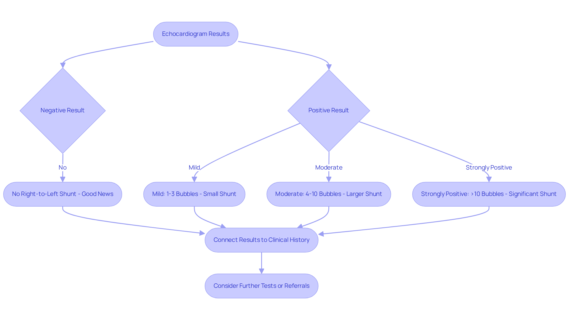
Negative Result: If no air pockets are detected in the left atrium, it usually means there’s no right-to-left shunt present. This is good news, as it suggests your heart is functioning normally in this regard.
-
Positive Result: If bubbles are found in the left atrium, it indicates a shunt and may necessitate an intervention. Here’s how we classify the degree of positivity:
- Mild: A small number of bubbles (1-3) indicate a minor shunt.
- Moderate: A moderate number of bubbles (4-10) indicate a larger shunt.
- Strongly Positive: More than 10 bubbles point to a significant shunt that may need treatment.
-
It’s essential to connect these results from the echocardiogram to your clinical history to create a tailored management plan. For example, in a study involving 880 patients treated for cryptogenic stroke, the presence of a right-to-left shunt was associated with a higher risk of recurrent stroke. This highlights the importance of thorough evaluation.
If your results indicate a serious condition, such as stroke, don’t hesitate to reach out for further tests or referrals to specialists. Remember, you’re not alone in this journey, and there are resources available to support you.
Conclusion
Echocardiograms, especially the specialized bubble study, are vital tools for assessing heart health and diagnosing potential cardiovascular issues. By using ultrasound technology to visualize the heart’s structure and function, these tests offer invaluable insights that can guide treatment decisions and enhance patient outcomes.
In this guide, we’ve explored key aspects of echocardiograms and bubble studies, including their definitions, indications, procedural steps, and how to interpret results. These assessments play a crucial role in identifying conditions like patent foramen ovale and other shunts, which, if overlooked, could lead to serious health complications. It’s also important to carefully select patients for these procedures to ensure effective and efficient healthcare delivery.
Ultimately, understanding echocardiograms with bubble studies empowers you to take proactive steps in managing your heart health. By recognizing the value of these diagnostic tools, you can engage more meaningfully with your healthcare providers and advocate for the evaluations you need. Seeking timely assessments and following established guidelines can lead to better health outcomes and a clearer path to maintaining cardiovascular wellness.
So, if you have concerns about your heart health, don’t hesitate to reach out for support. Remember, you’re not alone on this journey, and taking these steps can make a significant difference in your overall well-being.
Frequently Asked Questions
What is an echocardiogram?
An echocardiogram is a gentle, non-invasive ultrasound examination that uses high-frequency sound waves to create dynamic images of the heart, helping to assess its structure and function.
What can echocardiograms help assess?
Echocardiograms help assess the size of the heart chambers, the condition of the valves, and the overall pumping efficiency of the heart.
What is a bubble study in echocardiography?
A bubble study is a specialized type of echocardiogram where a saline solution mixed with air is injected into a vein to create tiny microbubbles, enhancing the visualization of blood circulation.
Why are bubble studies important?
Bubble studies are particularly helpful in detecting irregularities such as shunts between heart chambers and are effective for diagnosing conditions like patent foramen ovale (PFO) and other structural heart defects.
How common are contrast echocardiograms in clinical practice?
Contrast echocardiograms are performed in about 6% of echocardiographic examinations, highlighting their growing importance in clinical practice.
What role did contrast echocardiograms play in stroke diagnosis?
In a study involving individuals who experienced acute ischemic strokes, contrast examinations were vital in diagnosing PFOs, identifying 54 cases, which underscores their significance in managing stroke care.
What is Amavita Heart and Vascular Health®’s approach to cardiovascular challenges?
Amavita Heart and Vascular Health® focuses on comprehensive cardiac evaluations and preventive strategies tailored to individuals’ lives, using advanced risk assessment tools and personalized interventions.
How does ongoing research impact echocardiograms and cardiovascular health?
Ongoing research aims to enhance the effectiveness of echocardiograms and contrast examinations in promoting cardiovascular health, with a commitment to advancing diagnostic methods and improving care.
List of Sources
- Define Echocardiograms and Bubble Studies
- echonous.com (https://echonous.com/category/case-studies)
- pmc.ncbi.nlm.nih.gov (https://pmc.ncbi.nlm.nih.gov/articles/PMC11546412)
- cardioserv.net (https://cardioserv.net/indications-echo-bubble-study)
- health.harvard.edu (https://health.harvard.edu/heart-health/what-is-a-bubble-study)
- pmc.ncbi.nlm.nih.gov (https://pmc.ncbi.nlm.nih.gov/articles/PMC7527391)
- Identify Indications for Echo Bubble Studies
- pmc.ncbi.nlm.nih.gov (https://pmc.ncbi.nlm.nih.gov/articles/PMC11546412)
- pubmed.ncbi.nlm.nih.gov (https://pubmed.ncbi.nlm.nih.gov/37734655)
- cardioserv.net (https://cardioserv.net/indications-echo-bubble-study)
- Explain the Procedure for Conducting an Echo Bubble Study
- cardioserv.net (https://cardioserv.net/9-steps-to-perform-an-echo-bubble-study)
- aapc.com (https://aapc.com/codes/coding-newsletters/my-cardiology-coding-alert/you-be-the-coder-master-the-bubble-study-153124-article?srsltid=AfmBOop2ys0qNuNvXiwtsCs6O30BqaiHg9zEqH-jQxqQT67zXS3hJGow)
- Interpret Results of Echo Bubble Studies
- memorialcardiology.com (https://memorialcardiology.com/how-does-a-positive-bubble-study-work)
- cardioserv.net (https://cardioserv.net/indications-echo-bubble-study)
- sciencedirect.com (https://sciencedirect.com/science/article/pii/S0741521409016218)
- sciencedirect.com (https://sciencedirect.com/science/article/pii/S1936879818307167)
- researchgate.net (https://researchgate.net/publication/393009582_Bubble_Enhanced_Echocardiography_Practical_Insights)